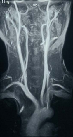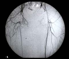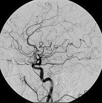
Angiography
Performing angiography can help to diagnose many conditions of the arteries, but the main disease studied by angiography is atherosclerosis, or blockages in the arteries. In the heart, blockages can cause heart attacks. In the brain, blockages can cause stroke. In the kidneys, blockages can cause high blood pressure and poor renal function. In the legs, blockages can cause leg pain or non-healing skin ulcerations.
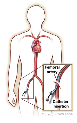
The catheters (tubes) used in angiography are very small, about the size of the tip of a pencil. They make a small hole in the artery in the hip area, which heals with gentle pressure applied after the procedure, and a few hours of bedrest. The x-ray dye used in modern angiography is very gentle on the body, now causing minimal discomfort and minimal effect on the kidneys, in small doses. VITA physicians always attempt to use as little as possible, only the amount to diagnose and treat artery conditions.
VITA physicians are board certified in the performance and interpretation of angiography, having completed many years of training and recertification in these techniques.
- For more information, please visit:
http://www.sirweb.org/patients/angiography/